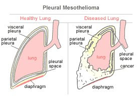
Pleural mesothelioma is the most common type of mesothelioma, a rare cancer that develops in the mesothelial cells that make up the mesothelium, a membrane that lines many of the body’s organs and cavities. In the case of pleural mesothelioma, the cancer develops in the lining of the lungs, called the pleura or pleural membrane.
The pleura is comprised of two layers which provide support and protection for the lungs and chest cavity. The outer layer, or the parietal layer, lines the entire chest cavity and the diaphragm. The inner layer, or visceral layer, covers the lungs. Fluid between these two membranes allows them to slip against one another as the lungs expand and contract. Pleural mesothelioma typically develops in one layer, but can metastasize, or spread, to the other layer.
Sometimes doctors refer to this disease as mesothelioma of the pleura. Mesothelioma is a cancer of the serous membranes. These membranes enclose a number of organs throughout the midsection of the body, including the lungs. The most common type of mesothelioma, pleural mesothelioma, affects the serous membranes of the lungs.
When mesothelioma spreads to the lungs from the serous linings of the lungs, abdomen or heart, it is considered secondary lung cancer. Also, pleural mesothelioma is sometimes referred to as an asbestos lung cancer. Technically, cancers that do not originate in the lungs are not considered lung cancer; thus, terms such as secondary lung cancer and asbestos lung cancer (pleural mesothelioma) are misleading. Asbestosis is a type of asbestos lung disease that does originate in the lungs and is often confused with mesothelioma.
Like all mesothelioma cancers, pleural mesothelioma is caused by asbestos exposure and develops when the toxic asbestos fibers become trapped in the spaces between the mesothelial cells.
Pleural Mesothelioma Symptoms
Pleural mesothelioma patients display all three types of mesothelioma cancer cells: epithelioid mesothelioma, sarcomatoid mesothelioma and biphasic mesothelioma.
Once trapped in the body, asbestos fibers cause cancerous cells to divide abnormally, resulting in the thickening of the pleural membrane layers and mesothelial cells, causing build-up of fluid (called pleural effusion). The fluid begins to put pressure on the lungs and the respiratory system in general, preventing normal breathing.
Symptoms of pleural mesothelioma are largely caused by these developments and may include the following:
- Persistent dry or raspy cough
- Coughing up blood (hemoptysis)
- Difficulty in swallowing (dysphagia)
- Breathlessness (dyspnea)
Shortness of breath that occurs even when at rest. Along with shortness of breath, patients may suffer from a cough. Rarely, patients may develop hoarsness or cough up blood (hemoptysis).
- Chest pain
Chest pain is often nonspecific, and may sometimes be felt in upper abdomen, shoulder, or arm. Chest pain and breathlessness are the most common, and usually earliest presenting, symptoms of pleural mesothelioma. Persistent pain in the chest or rib area, or painful breathing.
- Pleural effusion
A pleural effusion is the result of too much fluid building up between the parietal and visceral pleura (linings of the chest and lungs, respectively); a pleural effusion may cause chest pain and difficulty breathing (dyspnea), however, many cause no symptoms and are first discovered during the physical examination or seen on a chest x-ray.
- Development of lumps under the skin on the chest
- Night sweats or fever
Less common, but still cited enough to be considerd a symptom of pleural mesothelioma are fever, chills, and night sweats.
- Weight Loss
Unexplained weight loss is cited as a symptom in about a third of pleural mesothelioma cases.
- Fatigue
Pleural Mesothelioma Diagnosis
As with other types of mesothelioma, pleural mesothelioma is difficult to diagnose since symptoms do not typically arise for some time after initial asbestos exposure occurs. Additionally, since the symptoms of pleural mesothelioma are typical of many illnesses, in the early stages of the cancer the symptoms are often mistaken for less threatening diseases such as influenza and pneumonia.
X-rays or CT-Scans are often used to diagnose pleural mesothelioma.
A pleural mesothelioma diagnosis is made partly on the basis of symptoms but additional diagnostic tests are needed to confirm the presence of cancer. Following a medical history review and physical examination, patients must typically undergo imaging tests, such as x-rays or CT scans, to confirm the location of cancer. A patient must also usually endure fluid and tissue tests, also known as biopsies, to confirm the type of cancer involved.
Pleural effusions and peritoneal effusions are experienced by two-thirds of patients. Hemothorax - the collection of blood in the pleural cavity - also is a symptom. To get a diagnosis, doctors use imaging technologies as well as histological analysis and molecular biologic analyses. A pleural smear examines a sample of pleural fluid under the microscope to detect for abnormal organisms. The test is performed when infection of the pleural space is suspected or when an abnormal collection of pleural fluid is noticed by chest X-ray. Sometimes the tumor grows through the diaphragm, making the site of origin difficult to assess.

Top left, A: posterior-anterior chest radiograph in a patient with malignant pleural mesothelioma demonstrating significant right-sided pleural effusion and diffuse pleural thickening associated with marked volume loss of the right hemithorax. No definite pleural plaques are seen. Top right, B: CT image from a patient with a right-sided pleural mesothelioma, illustrating complete encasement of the ipsilateral lung with a thick rind of tumor, neoplastic invasion of the interlobar fissures, small residual pleural effusion, and marked unilateral volume loss. Center, C: MRI with transaxial, sagittal, and coronal views from a patient with a right-sided pleural mesothelioma. MRI may be most beneficial in the determination of chest wall, mediastinal, and diaphragmatic invasion. Bottom, D: PET scan with 18-FDG in the same patient as in center panel (C) with transaxial, sagittal, and coronal views. 18-FDG PET scanning in mesothelioma can aid in tumor staging, localization of biopsy sites, and distinguishing between pleural fibrosis and active malignant tissue. (resource : chest journal)
Pleural Mesothelioma Treatments
In general, pleural mesothelioma patients have three options: surgery, chemotherapy and radiation therapy. Typically, patients will receive a combination of two or more of these types of treatment.
Read More about Pleural Mesothelioma Treatments
Asbestos: The Primary Cause
Inhaling asbestos fibers is the primary cause of pleural mesothelioma. Other factors such as genetics, smoking, and exposure to the simian virus (which contaminated some older polio vaccines) may predispose some people to develop mesothelioma when exposed to asbestos. Some individuals exposed to asbestos for a prolonged period do not develop mesothelioma or asbestosis.
Asbestos is a naturally occurring mineral that was widely used in various industries and construction because of its fire-retarding and insulating properties. Since the 1980s, its use in industry and consumer products has been more regulated and some U.S. legislators would like to ban it entirely.
Serpentine and amphiboles are the two main forms of asbestos. Chrysotile asbestos, which is most commonly used in commercial products, is the only type of serpentine. Amphiboles are divided into five main types: actinolite, amosite, anthrophyllite, crocidolite, and tremolite.
Serpentine fibers are flexible and curly. Amphiboles have long, thin fibers and are thought to be a greater cancer risk than serpentine fibers, but exposure to either serpentine or amphibole asbestos is dangerous.
How Asbestos Exposure Occurs
People who worked for an extended period of time in an environment where asbestos dust was regularly released are at greatest risk for pleural mesothelioma. The body's natural defense mechanisms successfully clear many asbestos fibers, but when asbestos exposure is heavy, these defense systems are overwhelmed. Mucus within the airways traps asbestos fibers and other toxins that are inhaled. These trapped foreign particles are either swallowed or coughed up.
These fibers travel to the lungs and become imbedded in the lung lining, outside of the lungs and inside the ribs. These long, pointed fibers can reach the pleural lining of the chest wall and lung. Once the fibers have penetrated the pleura they injure the mesothelial cells. When these jagged particles settle in the pleura, they cause inflammation. The inflammation, in turn, can lead to dangerous cancerous tumors. In some cases, those who've inhaled asbestos fibers will first develop the less-severe asbestosis, followed by mesothelioma several years later.
Upon diagnosis, patients usually exhibit multiple tumor masses affecting both the visceral (further from the lung) and parietal surfaces (closer to the lung) of the pleura. The parietal surface is more often affected than the visceral surface, and the right lung, due to its larger size, often suffers more damage than the smaller left lung. In addition, more asbestos tends to settle in the lower lungs than the upper lungs.
These tumors often grow quickly in size and can cover the entire lung cavity, making it very difficult to breathe and causing excruciating pain. Also, in the advanced stages of pleural mesothelioma, the cancer may spread to other nearby organs, including the heart, abdomen, and lymph nodes.
The tissue changes leading to mesothelioma occur slowly. The symptoms of mesothelioma generally do not appear for 20 to 30 years after the initial asbestos exposure. A latency period of up to 50 years has been known.
resource :
- allaboutmalignantmesothelioma
- asbestos.com
- pleuralmesothelioma.com


